
The airway anatomy is a vital part of your body, and its proper functioning is of the utmost importance. It consists of the nose and lungs, obviously. But do you know what the pharynx does?
Below, we talk about the structure and function of various parts of your airway anatomy — AKA, your respiratory tract.
But it’s also important to know the difference between child and adult airway anatomy. We provide for you an easy-to-read bullet list of differences that every parent should learn.
Proper airway health can mean better sleep, less fatigue, and even better oral health. Poor airway health can lead to sleep apnea, infection, and worse.
What is the airway?
The airway is your respiratory tract, the part of your body that controls breathing. An airway refers to your breathing passages, through which oxygen is taken in and carbon dioxide is expelled.
Your airway starts with your nose (or mouth), continues through your windpipe (trachea), and ends in your lungs, where alveoli give your blood oxygen and extract carbon dioxide.
The epithelium (outer layer) of your airway is mostly lined with mucous membranes to moisten airflow and prevent pathogens from getting in, as well as hairlike cilia which superiorly move debris or mucus and sense the direction of airflow.
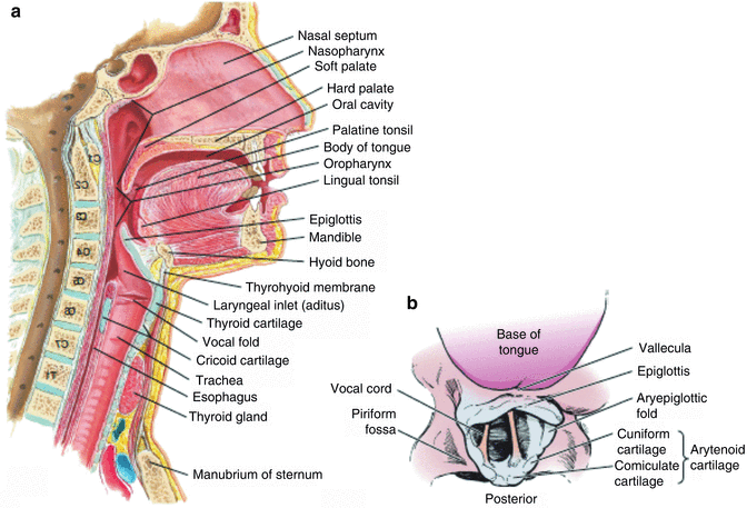
What is the anatomy of the airway? The anatomy of the airway begins with the nose and nasal cavity (or oral cavity), then proceeds through the pharynx, past the epiglottis, into the larynx where your vocal cords are, down the trachea, through the bronchial tree, and into the lungs.
Upper Airway Anatomy
The upper airway consists of:
- Nose — This is the main entryway to your respiratory system. The mucosa of the nose filters, warms, and moistens airflow as it passes through. It allows you to smell and even taste.
- Nasal cavity — Divided into 2 nasal passages continuing from both nostrils (nares), the nasal cavity also filters and warms the air passed through it, headed for the pharynx.
- Paranasal sinuses — These surround and drain into the nasal cavity. Their main purposes of the paranasal sinuses is debated.
- Pharynx — This connects the nasal cavity and oral cavity to the trachea and esophagus. The pharynx can be broken into 3 distinct portions: the nasopharynx, the oropharynx, and the laryngopharynx.
The upper respiratory tract can also be said to include the part of the larynx above the vocal cords, but some experts simply group the larynx in with lower airway anatomy.
What separates the upper and lower airway? The epiglottis separates the upper and lower airways. Located just above the glottis (the top portion of the larynx), the epiglottis opens posteriorly to allow breathing into the trachea and closes to allow swallowing down the parallel esophagus.
Lower Airway Anatomy
The lower airway, or lower respiratory tract, consists of the following organs:
- Larynx — Located inferior to the hyoid bone, this houses the vocal folds (vocal cords), which vibrate to make sounds, open to allow breathing, and close when speaking or swallowing. The laryngeal reflex is considered one of the strongest human reflexes.
- Trachea — Also known as the windpipe, this provides a passage for oxygen to travel down (and carbon dioxide to travel up), while warming and moistening the air as it passes through. The trachea is the most anterior (in front) part of the neck, except when the thyroid covers it briefly. Located midline or perhaps slightly deviated to the right due to the heart’s placement, the trachea extends from the neck to the thorax.
- Bronchial tree — At the bottom of the trachea, branches of bronchi (e.g., right bronchus or mainstem bronchi) and smaller bronchioles extend right and left to distribute air and oxygen to the lungs. These branches of bronchi are called the bronchial tree. The intersection of these branches with the trachea is called the carina.
- Lungs — These bring fresh oxygen into your body by expanding and remove carbon dioxide and other waste gases by contracting. The expansion and contraction is possible due to the diaphragm, as well as the external intercostal muscles between the ribs. You have two of these: right lung and left lung, both in your thoracic cavity.
- Alveoli — These are a part of the lungs. Alveolar ducts and sacs are responsible for the gas exchange of oxygen and carbon dioxide. Alveolar sacs and capillaries form the air-blood barrier.
The lower airway possesses a layer of smooth muscle allowing precise control over bronchoconstriction.
Is the larynx the upper or lower airway? The larynx is part of the lower airway. The upper airway includes the similarly named pharynx.
Child vs. Adult Airway: Differences and Development
Here are some of the main differences between child airways and adult airways:
- The child’s airway is smaller in diameter and shorter in length than the adult’s airway. Even minor injuries or slight swelling can make it harder for a young child to breathe.
- The pediatric larynx is higher and located more anteriorly than the adult’s larynx.
- A younger child’s tongue is relatively larger within the oropharynx than an adult’s tongue.
- Infants are nose breathers only for the first 4-6 months of life.
- A child’s trachea is softer, consisting more of cartilage than an adult’s. This increases the risk of windpipe collapse or obstruction in children.
- The pediatric trachea and neck are shorter and more cartilaginous than in adults. The pediatric windpipe begins at the 3rd or 4th cervical vertebrae, whereas the adult’s starts at the 5th or 6th cervical vertebrae.
- The child’s occipital bone (the back and lower part of the skull) is large and round, compared to the flatter adult skull.
- Enlarged tonsils and/or adenoids are thought to be the leading cause of obstructive sleep apnea in children. For adults, enlarged tonsils and/or adenoids are much less likely to obstruct the upper airway.
- The adult epiglottis is flat, flexible, and the narrowest part of the airway. The child’s epiglottis is horseshoe-shaped, shorter, and stiffer. And in a child’s airway, the cricoid cartilage is thought to be the narrowest region.
Common Airway Problems
Below are 8 common respiratory and airway problems:
- Airway obstruction
- Asthma
- Bronchitis
- Chronic obstructive pulmonary disease (COPD)
- Cystic fibrosis
- Emphysema
- Pneumonia
- Sleep apnea
Airway obstruction, whether chronic or acute, may be caused by:
- small object lodged in throat
- sleep apnea
- large tongue
- swollen tonsils and/or adenoids
- swollen epiglottis
- infection
- trauma or injury
- smoke inhalation
- allergies
- asthma
- chronic bronchitis
- chronic obstructive pulmonary disease (COPD)
- croup
- cystic fibrosis
- emphysema
What is “airway health”?
Airway health is the idea that your airway’s well being affects the health of other parts of the body.
If you have an airway obstruction, this can cause teeth grinding (bruxism) which is horrible for your dental health. Or if you don’t get enough oxygen due to poor airway health, this reduces your energy and may lead to headaches and even confusion.
At Rejuvenation Dentistry, we know how important airway health is. Our founder, Dr. Gerry Curatola, has decades of experience identifying and treating sleep apnea (a major airway health issue) with the DNA Appliance or other biological dentistry methods.
Mallampati Scores
Mallampati scores identify the size of your tongue relative to your pharynx, on a scale of 1 to 4.
Also called the Mallampati classification, this is a simple test that helps predict ease of intubation as well as an obstructive sleep apnea diagnosis.
Here is an easy visualization of what it looks like to score a 1, 2, 3, or 4 — also referred to as Class I, II, III, and IV.
https://www.instagram.com/p/B_Z546vJsPF/
In a sitting position, a healthcare professional will ask the patient to open the mouth and protrude the tongue as much as possible. Depending on if the base of the uvula, faucial pillars, and soft palate are visible, scores are given as follows:
- Class I: soft palate, all of uvula, and faucial pillars visible
- Class II: soft palate, most of uvula, and faucial pillars visible
- Class III: soft palate and base of uvula visible
- Class IV: only hard palate visible
Benefits of a Healthy Airway
A healthy airway means good sleep, less fatigue, and better overall health.
If you’re worried about sleep apnea, you’re not alone. Conventional doctors don’t address the root cause of obstructive sleep apnea — your tongue is too large for your oral cavity.
Rejuvenation Dentistry makes this their problem and aims to solve it with the DNA Appliance and other conservative biological dentistry techniques.
Did you know? A landmark 2006 study on 145,000 patients found up to a 21% reduction in healthcare costs associated with major diseases when oral health is improved.
Set up your appointment with us right away. At Rejuvenation Dentistry, we empower our patients to take control of their overall health, including airway health problems and obstructive sleep apnea.
Sources
- Harless, J., Ramaiah, R., & Bhananker, S. M. (2014). Pediatric airway management. International journal of critical illness and injury science, 4(1), 65. Full text: https://www.ncbi.nlm.nih.gov/pmc/articles/PMC3982373/
- Kostrzewa-Janicka, J., Jurkowski, P., Zycinska, K., Przybyłowska, D., & Mierzwińska-Nastalska, E. (2015). Sleep-related breathing disorders and bruxism. In Ventilatory Disorders (pp. 9-14). Springer, Cham. Abstract: https://pubmed.ncbi.nlm.nih.gov/26022906/
- Liistro, G., Rombaux, P. H., Belge, C., Dury, M., Aubert, G., & Rodenstein, D. O. (2003). High Mallampati score and nasal obstruction are associated risk factors for obstructive sleep apnoea. European Respiratory Journal, 21(2), 248-252. Full text: https://erj.ersjournals.com/content/21/2/248
- Albert, D. A., Sadowsky, D., Papapanou, P., Conicella, M. L., & Ward, A. (2006). An examination of periodontal treatment and per member per month (PMPM) medical costs in an insured population. BMC Health Services Research, 6(1), 103. Full text: https://www.researchgate.net/publication/6873716_An_examination_of_periodontal_treatment_and_per_member_per_month_PMPM_medical_costs_in_an_insured_population