Cone beam computed tomography (cone beam CT, or CBCT) is an advanced x-ray that allows clinicians to take a 3D image of the mouth. Unlike a standard dental x-ray, cone beam CT also images the soft tissues of the mouth, including the gums, tongue, and nerves.
Dental cone beam computed tomography is a non-invasive way for dental professionals to get a complete picture of a patient’s mouth. It exposes patients to substantially less radiation than a traditional CT, making it a top choice for biological dentists who need detailed imaging.
What is cone beam in radiology? Cone beam in radiology is a radiographic imaging device that focuses an x-ray beam into a cone shape. That beam is used to create a 3D image of a body region, usually the mouth or head.
History Of The Cone Beam CT
The cone beam CT was concurrently developed by Dr. Piero Mozzo and Dr. Yoshinoro Arai in the mid-1990s. The doctors created the cone beam CT to improve upon the traditional CT scanner and create a 3D-imaging device that worked better for dental imaging.
How does a CT scan work? CT scans work by sending high-energy radiation through the body to a detector, taking multiple images. A computer connected to the CT system then uses an algorithm to put the images into a 3D model of the scanned area.
Cone Beam CT Vs. Conventional CT
What is the difference between CT and CBCT? The difference between CT and CBCT is how the x-rays are sent out from the x-ray source. Traditional CT uses a fan-shaped beam or spiral scan for medical imaging, but CBCT uses a cone-shaped beam, as the name suggests.
Conventional CT scanners use anode x-ray tubes to produce the radiation that’s used to perform imaging. Cone beam CT scanners, on the other hand, use a medical fluoroscopy tube, which generates less radiation but still captures incredibly high-resolution radiographs.
Both cone beam CT and conventional CT scanners use a detector to collect any x-rays that pass through the patient’s body. CBCT technology then converts the data from the detector into voxels, essentially 3D pixels, which create a digital reconstruction of the area.
Cone beam CT scanners are also smaller and less expensive to purchase, making it easier for a dental practice to have one in their offices. Conventional CT scanners are too large and cost significantly more, which is why they’re typically found only in imaging centers.
Common Uses Of A Cone Beam CT
The American Academy of Oral and Maxillofacial Radiology (AAOMR) recommends the use of CBCT to image the maxillofacial region before making bone grafts or installing dental implants. Having a detailed 3D image helps your oral surgeon with implant planning.
Cone beam CT scans can also be used after implants have been placed to monitor how the implant is integrating with the bone of the jaw. It provides more detail than a standard x-ray, and the 3D images can give dentists a better idea of how osseointegration is progressing.
CBCT imaging can be used to diagnose mouth and jaw issues such as dental cavitations. It also helps dental professionals investigate problems with the nerve canals of the jaw and other issues arising from changes to the structure of the jaw.
Additionally, CBCT scans are used in orthodontics and endodontics. Cone beam CT scans provide much higher resolution than other dental radiography modalities like bitewing or panoramic x-rays. This, in turn, improves treatment planning and results for procedures like root canals.
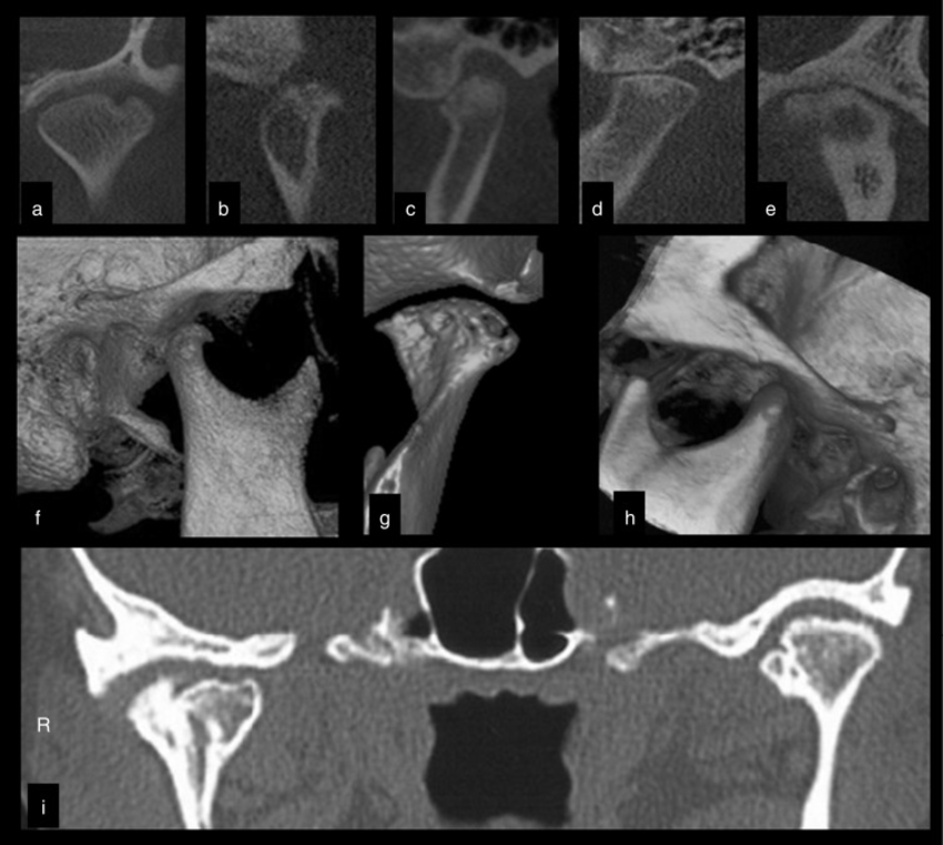
Cone beam CT imaging is commonly used to get a complete visualization of the temporomandibular joint (TMJ) in patients with jaw pain. It is one of the best ways to get a high-resolution look at any changes in the bony structures in and around the jaw joint.
Cone beam CT has clinical applications outside of dental practices as well. Otolaryngologists (ENTs) may also use CBCT systems to image the sinuses. CBCT can pick up on changes in the soft tissues of the sinuses that aren’t visible in x-rays.
What is cone beam CT used for? Cone beam CT is used to create high-resolution 3D images of the body region that’s scanned. It’s most often used in dentistry and oromaxillofacial applications, but can be used to radiographically image many parts of the body.
Potential Cone Beam CT Benefits
There are many benefits of using dental cone beam CT, especially when compared to a traditional CT scan. Many practitioners, dentists included, are switching over to using cone beam CT because it’s superior to other types of imaging.
What are the benefits of CBCT? The benefits of CBCT are:
- Lower radiation exposure compared to conventional CT scans
- Higher image quality and greater detail of CBCT images, particularly compared to standard x-rays
- The 3D image produced by CBCT gives your clinician more information than an x-ray
- CBCT images both soft tissue and bone, unlike x-rays
- Faster scan times because everything is scanned in a single rotation
- CBCT is totally painless and noninvasive
Is cone beam CT safe? Yes, a cone beam CT is safe. It may be safer than a conventional CT scan in most cases. A conventional CT exposes a patient to 10 times the radiation dose of a cone beam CT.
Possible Complications Of Cone Beam CT Use
Although cone beam CT uses less radiation than a conventional CT scan, it still exposes patients to more radiation than a simple x-ray. High doses of radiation can eventually lead to cancer, although a single cone beam CT scan doesn’t come close to putting off that level of radiation.
Dentists only use cone beam CT when they can’t get the information they need from traditional x-rays. In some instances, dentists need 3D visualization or high-resolution images that x-rays can’t give them.
Radiologists, dentists, and other medical professionals follow ALARA. ALARA standards mean they keep radiation exposure As Low As Reasonably Achievable. In many cases, cone beam CT provides images with the best detail and the lowest dose of radiation possible.
Children are particularly sensitive to radiation and should only receive a cone beam CT or conventional CT scan in sporadic cases when the scan is essential. In a dental practice, these would probably be cases in which a decision must be made about how to treat a cavity.
Pregnant women should also avoid CT scans unless there’s a strong medical need to have one.
What To Expect In A Cone Beam CT Scan
Preparing for a cone beam CT scan is quite easy. You’ll be asked to remove any glasses, hearing aids, jewelry, and metal hair accessories, so you may want to plan accordingly. It’s also best to wear comfortable clothing and shoes if setting up the machine takes a few minutes.
Women who are pregnant or might be pregnant should tell their doctor before having a cone beam CT scan. The higher levels of radiation could be harmful to the fetus. Your doctor will decide whether the benefits of a CBCT scan outweigh the risks.
Getting a dental CBCT scan is very similar to having panoramic x-rays of your teeth taken. You’ll stand or sit in the middle of the machine and gently bite on a piece of plastic to position your teeth properly.
Your dentist or radiologist may also add straps to hold your head in the right place. It’s vital to stay as still as possible, so you get the sharpest picture with the greatest detail. The straps make it easier for you to keep your head from moving.
The initial setup may take several minutes, but the scan itself takes just a few seconds, generally 40 seconds at most. Your scan won’t make a lot of noise like an MRI machine, so there’s no need to worry if you’re sensitive to loud sounds.
During the scan, the CBCT machine will move around your head, like machines that take panoramic x-rays. You simply sit or stand still, depending on the machine, and let it do the work.
Do I need to be sedated to get a CBCT? No, you do not need to be sedated to get a CBCT. The process is completely painless, non-invasive, and only takes a few minutes.
The images from your cone beam CT scan will be sent to your dentist, oral surgeon, or radiologist. They will interpret the images and use them to make any diagnoses or treatment plans based on what they see.
Cone Beam CT Cost
The cost of a cone beam CT scan usually ranges between $100 and $600, depending on how much of your head needs to be imaged.
Your medical or dental insurance may cover some or all of the cost of imaging. However, it’s a good idea to check with your dental insurance provider beforehand if you’re unsure.
Connect With A Biological Dentist
Biological dentists are not just concerned with your teeth and mouth; they’re concerned about your overall health and wellbeing. They work to minimize your exposure to toxic chemicals and unnecessary radiation so you can stay healthy while protecting your oral health.
Rejuv has the best biological dentists in the country. Make an appointment to see us at our Manhattan or East Hampton offices to learn how we can keep your mouth healthy holistically.